
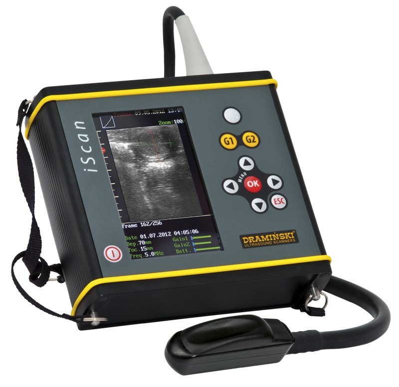
DRAMINSKI iSCAN Veterinary ultrasound scanners (with integrated linear, rectal probe, 5.0' LCD, LED display, 2 batteries, 1 shade cover)
Dimensions:17.0 x 17.0 x 6.0 cm
Unit weight:1720 g including probe
Battery weight::280 g
Image presentation (projection) mode:B Mode – live imaging,
B+B Mode (2B Mode – Dual Mode Display),
Probe frequency and type:electronic linear 7.5 MHz (from 4 to 9MHz)
Scanning range:from 4 to 12 cm (for linear probe, rectal use)
ScreenLCD, LED screen, 5.0’’ diagonal
Keyboard:membrane
Cineloop (image sequence):256 frames (ca. 20 seconds)
Image memory:200 images with date, index No. and description
Cine memory:50 cine loops with date, index No. and description
Transmission to the computer:through the USB2.0 port
Power source:Li-ion battery pack, 14.4 V, 3.1Ah
Continuous operation on fully charged battery:about 5 h
Pack charging time:2 hrs 30 min
Battery low indicator:automatic – graphic indicator
Working temperature:+ 5°C do + 40°C
Storage temperature:0°C do + 45°C
The set includes:
scanner’s body with electronic linear, rectal probe,
carrying strap, which allows hanging the device on the neck,
package of external batteries (1 item in a standard version, 2 items in prestige version),
battery charger,
power cable for the charger,
USB cable for data transmission,
shade cover
a bottle of ultrasound gel (in prestige version),
abridged manual,
manual and a programme for data transmission to a PC, recorded on a CD,
Explorer transport case.
A portable, fully digital ultrasound scanner with linear rectal probe. A reliable device for accurate diagnosis of, for instance, full breeding of cows and mares, tendons and horse’s equine eye ball, etc.
Ultrasonography is a dynamically developing and most popular method for imaging of organs and tissues in veterinary medicine. Miniaturisation of electronic systems and efficient power sources enabled Dramiński to design a truly revolutionary ultrasound model for field use. At the designing stage, we took into account most of the needs reported by veterinarians practising in difficult field conditions on a daily basis.
Why should you have DRAMIŃSKI iScan?
- Excellent image quality
A fully digital imaging technology has been used (B, B+B, B+M) to help in professional and fast imaging of:
- full breeding of cows and mares,
- tendons and horse’s equine eye ball,
- pathological mammary gland,
- objective assessment of condition of milk cows,
- other examinations conducted with the use of ultrasound technology, e.g. breeding in other animal species (fish, snakes, alpaca, camels)
- Small, light and mobile
By observing the physicians performing field work, we developed a design that involves:
- small weight of the device, which allows for long-term work in hard conditions,
- ultra-compact size to help examine without the assistance of other persons,
- very efficient and easy-to-replace external battery assures non-stop operation.
- Extremely durable
In the work of every physician devices need to be resistant to extremely harsh conditions. Therefore, the iScan ultrasound scanner:
- has a duraluminum, very strong casing,
- is fully resistant to water and other pollutants and can be washed under running water.
- High functionality
- intuitive membrane keyboard,
- an excellent LCD display with LED backlight, with a very wide viewing angle,
- a sun visor that enables the user to work even in full sun,
- clear and user-friendly menu,
- storage of ultrasound images and video loops (cine loop) in the device’s memory,
- transmission of saved images and cine loop to a computer via USB2.0.
Owing to the above featuresDramiński iScan gains very positive reviews around the world and is considered a model diagnostic device for work in very harsh conditions.
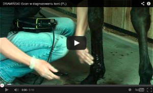
DRAMINSKI iScan the diagnosis of horses
DRAMINSKI iScan – waterproof test

Get exclusive volume discounts, bulk pricing updates, and new product alerts delivered directly to your inbox.
By subscribing, you agree to our Terms of Service and Privacy Policy.
Direct access to our certified experts